
FR, flexor retinaculum FDS, flexor digitorum superficialis tendons FDP, flexor digitorum profundus tendons MN, median nerve UN, ulnar nerve FCR, flexor carpi radialis tendon FPL, flexor pollicis longus tendon RA, radial artery UA, ulnar artery PQ, pronator quadratus muscle R, radius U, ulna PL, palmaris longus tendon R, radius U, ulna.įig. 3.1 Volar aspect of the wrist, axial ultrasound view. It may also vary in shape presenting different types of muscular and tendinous components as anatomic variants.įig. The palmaris longus tendon may be absent in up to 16% of the population as a normal variant. Outside the carpal tunnel are the tendons for the flexor carpi radialis, flexor carpi ulnaris and the palmaris longus. The bone margins of the tunnel, going from lateral to medial and proximal to distal are the scaphoid tubercle and pisiform bone and more distally, the ridge of the trapezium and the hook of the hamate all these bone margins are hyperechoic. The median nerve, a hyperechoic structure that contains small hypoechoic dots representing the neuronal fascicles these are best shown on transverse ultrasound view. Above the flexor tendons and below the flexor retinaculum is the median nerve. On ultrasound, all these tendinous structures show a hyperechoic fibrillar pattern. Deeper in this aspect is the carpal tunnel that contains the superficial and deep hand flexor tendons (flexor digitorum superficialis and profundus of the second to fifth finger) and their surrounding single synovial sheath, as well as the flexor pollicis longus tendon surrounded by its sheath. The volar aspect comprises a roof made by the flexor retinaculum, which is a fibrous band best identified on transverse view as a hyperechoic band. The wrist can be subdivided into four segments: the volar, dorsal, medial, and lateral aspects ( Figs. These flexo-extension maneuvers are performed along a longitudinal sonographic axis this dynamic study shows the normal and gentle movement of the tendons and can be helpful in the detection of tendon entrapments or for the identification of the median nerve inside the carpal tunnel that does not show any movement. Maneuvers of gentle flexion and/or extension of the digits can help identify the different tendons. For the examination of the volar aspect of the wrist, the hand is positioned palm up with mild extension and radial or ulnar deviation for examination of the dorsal aspect of the wrist, the hands are moved into a palm down position. In addition, oblique approaches or a heel-toe maneuver (to increase the pressure on one side of the probe) can be helpful to remove anisotropy artifacts. The screening on ultrasound examination includes transverse and longitudinal views of the local structures at rest or after dynamic testing positions. Alternatively, the patient may be seated at an examination table with both hands resting on the table. Space-occupying lesions-dorsal or ventral ganglia, neurogenic tumors, lipomatous tumors, presence of accessory musclesįor an ultrasound of the wrist, the patient rests the wrist in his or her lap and is seated in front of the examiner.

Traumatic lesions of extensor and flexor tendons, nerves, vessels, and occult fractures of the carpal bonesĮntrapment neuropathies-tunnel syndromes (carpal and Guyon) Inflammatory and degenerative diseases-De Quervain disease, distal intersection syndrome, tenosynovitis, Inflammatory arthritis (rheumatoid arthritis, crystal-related arthritis) Hence, ultrasound is the ideal imaging modality.
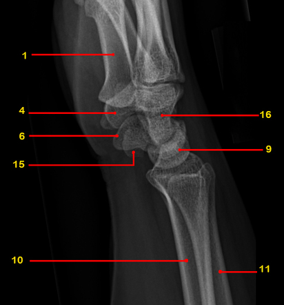
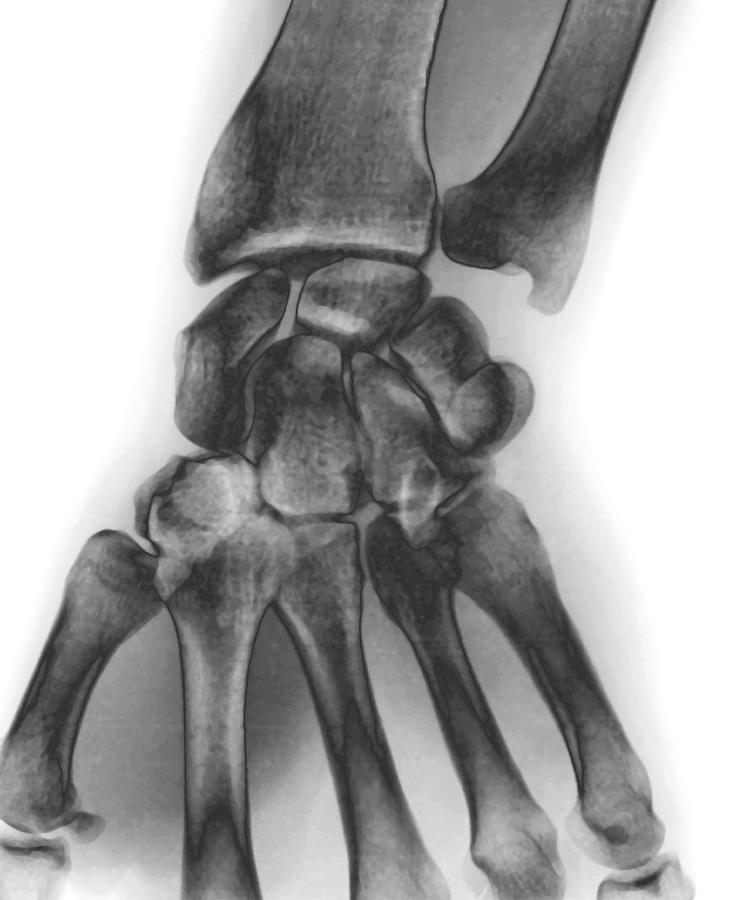
#Wrist xray normal skin
Pathology of the wrist includes both injuries and systemic diseases involving the wrist’s complex anatomy of bones, a network of soft tissues, and thin skin layers. Thus, ultrasound can be used for diagnostic purposes, for monitoring the results of treatments, or to guide percutaneous procedures.


When properly performed, ultrasound in addition to anatomic details provides functional data such as blood flow. For the wrist, which is easily accessible, the studies can be performed at rest or during dynamic testing as they are performed rapidly, without exposing the patient or the imager to ionizing radiation. The newest ultrasound systems capture high-definition images of tissues and lesions in real time.


 0 kommentar(er)
